Home

zaneprázdnený poľa viac lvidd echo normal spojenectvo orech chov

Introduction to Echocardiography, Parts 1-2 | IAME

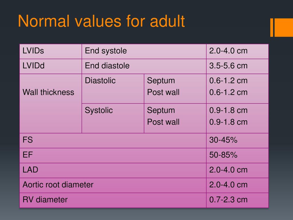
Echocardiography (Normal values) - TECHmED

Echocardiography (Normal values) - TECHmED

Introduction to Echocardiography, Parts 1-2 | IAME

Echocardiography (Normal values) - TECHmED

Cardiac Time Intervals by Tissue Doppler Imaging M-Mode: Normal Values and  Association with Established Echocardiographic and Invasive Measures of  Systolic and Diastolic Function | PLOS ONE
Cardiac Time Intervals by Tissue Doppler Imaging M-Mode: Normal Values and Association with Established Echocardiographic and Invasive Measures of Systolic and Diastolic Function | PLOS ONE

Should LV Mass be indexed with Height or BSA? Cardioserv

THE AMERICAN SOCIETY OF ECHOCARDIOGRAPHY RECOMMENDATIONS FOR CARDIAC  CHAMBER QUANTIFICATION IN ADULTS: A QUICK REFERENCE GUIDE F
THE AMERICAN SOCIETY OF ECHOCARDIOGRAPHY RECOMMENDATIONS FOR CARDIAC CHAMBER QUANTIFICATION IN ADULTS: A QUICK REFERENCE GUIDE F
Echocardiography: an overview - part I

Echocardiography for the Surgeons - ppt download

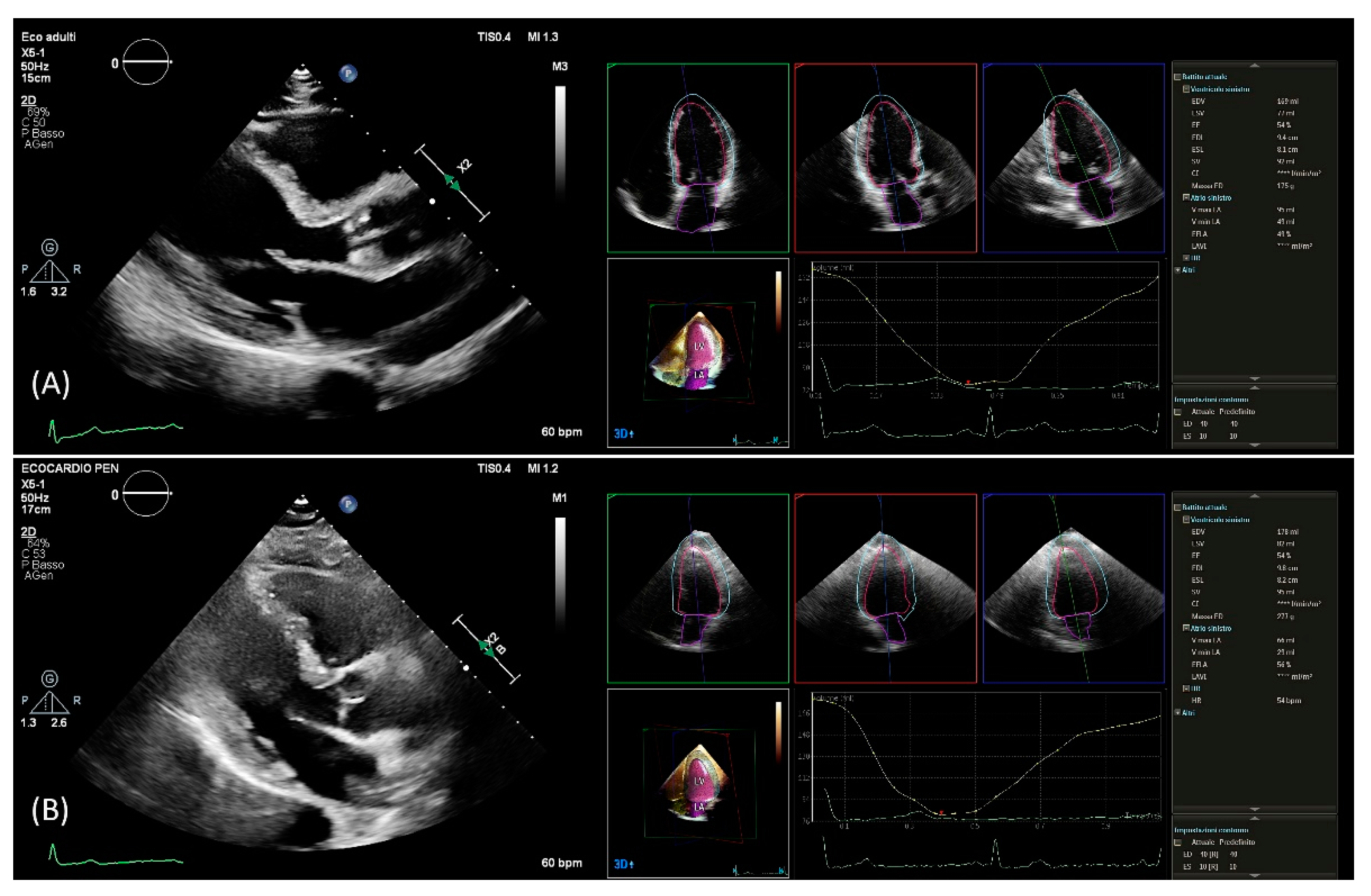
JCM | Free Full-Text | Echocardiographic Left Ventricular Mass Assessment:  Correlation between 2D-Derived Linear Dimensions and 3-Dimensional  Automated, Machine Learning-Based Methods in Unselected Patients
JCM | Free Full-Text | Echocardiographic Left Ventricular Mass Assessment: Correlation between 2D-Derived Linear Dimensions and 3-Dimensional Automated, Machine Learning-Based Methods in Unselected Patients

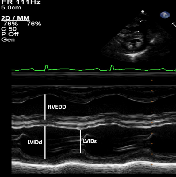
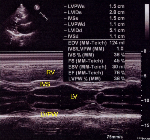
M-mode echocardiogram in left ventricular dysfunction – All About  Cardiovascular System and Disorders
M-mode echocardiogram in left ventricular dysfunction – All About Cardiovascular System and Disorders

M-Mode Echocardiographic Measurements – All About Cardiovascular System and  Disorders
M-Mode Echocardiographic Measurements – All About Cardiovascular System and Disorders

THE AMERICAN SOCIETY OF ECHOCARDIOGRAPHY RECOMMENDATIONS FOR CARDIAC  CHAMBER QUANTIFICATION IN ADULTS: A QUICK REFERENCE GUIDE F
THE AMERICAN SOCIETY OF ECHOCARDIOGRAPHY RECOMMENDATIONS FOR CARDIAC CHAMBER QUANTIFICATION IN ADULTS: A QUICK REFERENCE GUIDE F

Echocardiography in General Practice: Cardiac Abnormalities | Clinician's  Brief
Echocardiography in General Practice: Cardiac Abnormalities | Clinician's Brief

Routine Echocardiogram Protocol With Standard 2D Echo Images and Color  Doppler - YouTube
Routine Echocardiogram Protocol With Standard 2D Echo Images and Color Doppler - YouTube

Importance of length and external diameter in left ventricular geometry.  Normal values from the HUNT Study | Open Heart
Importance of length and external diameter in left ventricular geometry. Normal values from the HUNT Study | Open Heart

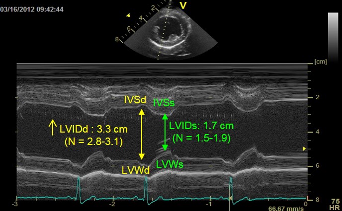
Measurement of normal LV diameter - UpToDate

The Left Ventricle and Its Systolic Function | Thoracic Key

Echocardiography (Normal values) - TECHmED

Normalized Echocardiographic Values From Guideline-Directed Dedicated Views  for Cardiac Dimensions and Left Ventricular Function | JACC: Cardiovascular  Imaging
Normalized Echocardiographic Values From Guideline-Directed Dedicated Views for Cardiac Dimensions and Left Ventricular Function | JACC: Cardiovascular Imaging

Left Ventricle
Left Ventricle

FUSIC Heart – FUSIC-SY

Difference between ECG and Echocardiography | Difference Between

Classification of left ventricular size: diameter or volume with contrast  echocardiography?
Classification of left ventricular size: diameter or volume with contrast echocardiography?