Home

primitívne debilné Prevádzka možná oval cross section dental obsluhovať Oženiť sa veľrybie

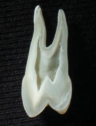
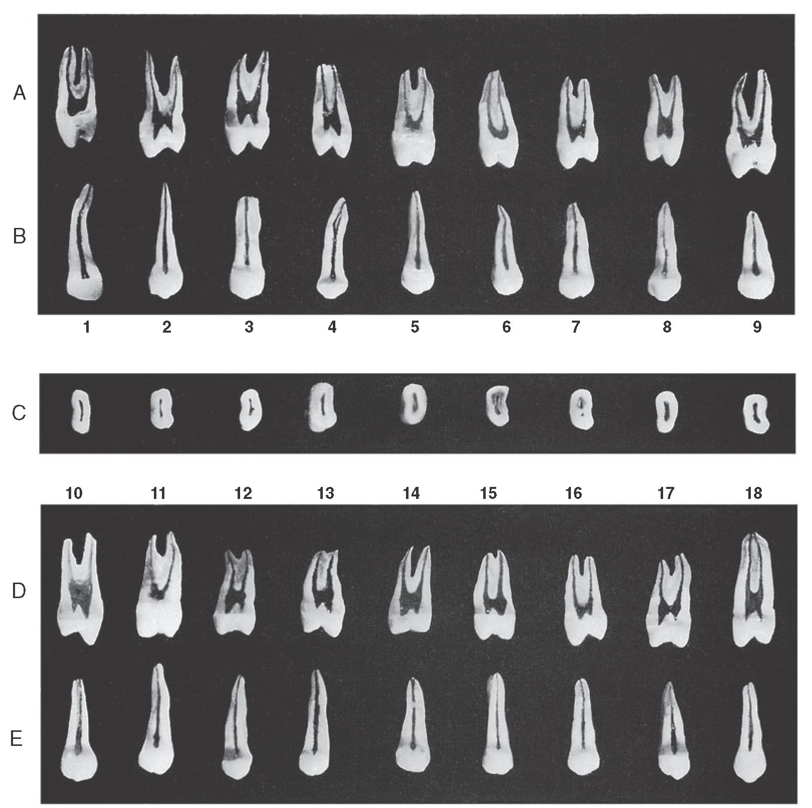
Pulp Chambers and Canals (Dental Anatomy, Physiology and Occlusion) Part 3

View Image
View Image

File:Cross sections of teeth labels.png - Wikimedia Commons

The oval shaped, root canal: A clinical review

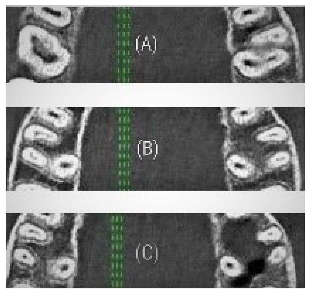
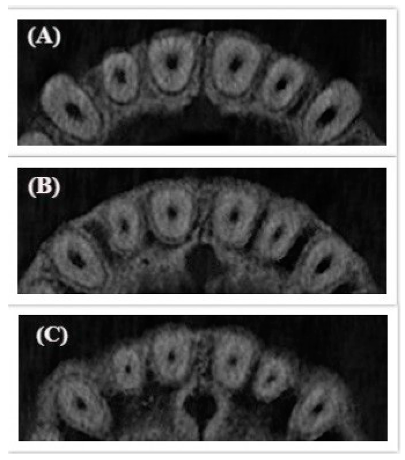
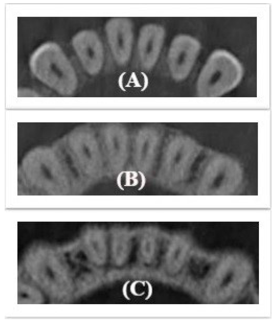
Applied Sciences | Free Full-Text | Evaluation of Cross-Sectional Root  Canal Shape and Presentation of New Classification of Its Changes Using  Cone-Beam Computed Tomography Scanning
Applied Sciences | Free Full-Text | Evaluation of Cross-Sectional Root Canal Shape and Presentation of New Classification of Its Changes Using Cone-Beam Computed Tomography Scanning
Root canal - Wikipedia

Applied Sciences | Free Full-Text | Evaluation of Cross-Sectional Root  Canal Shape and Presentation of New Classification of Its Changes Using  Cone-Beam Computed Tomography Scanning
Applied Sciences | Free Full-Text | Evaluation of Cross-Sectional Root Canal Shape and Presentation of New Classification of Its Changes Using Cone-Beam Computed Tomography Scanning
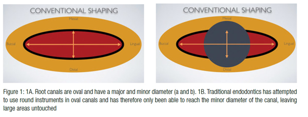
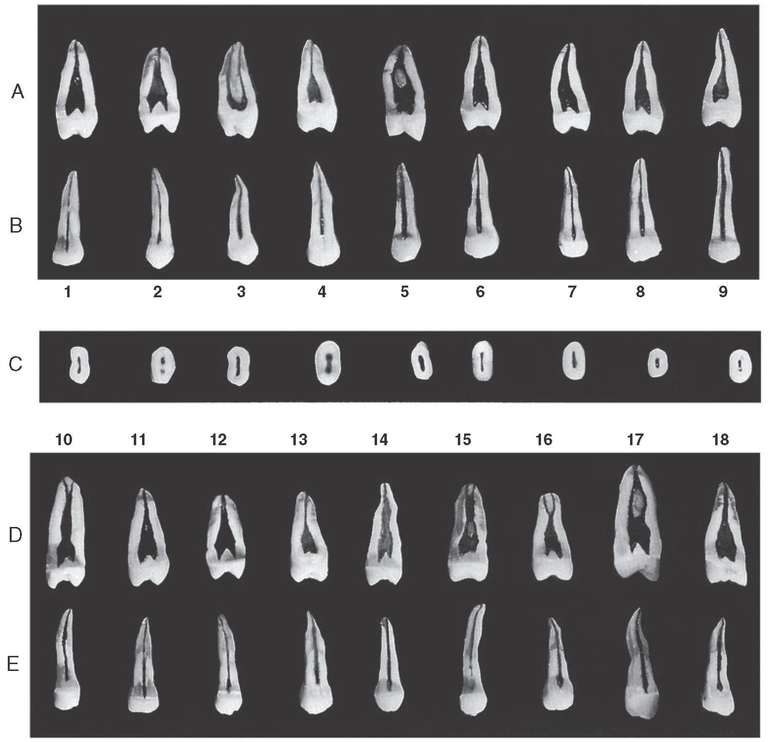
Endodontics: Part 4 Morphology of the root canal system | British Dental  Journal
Endodontics: Part 4 Morphology of the root canal system | British Dental Journal

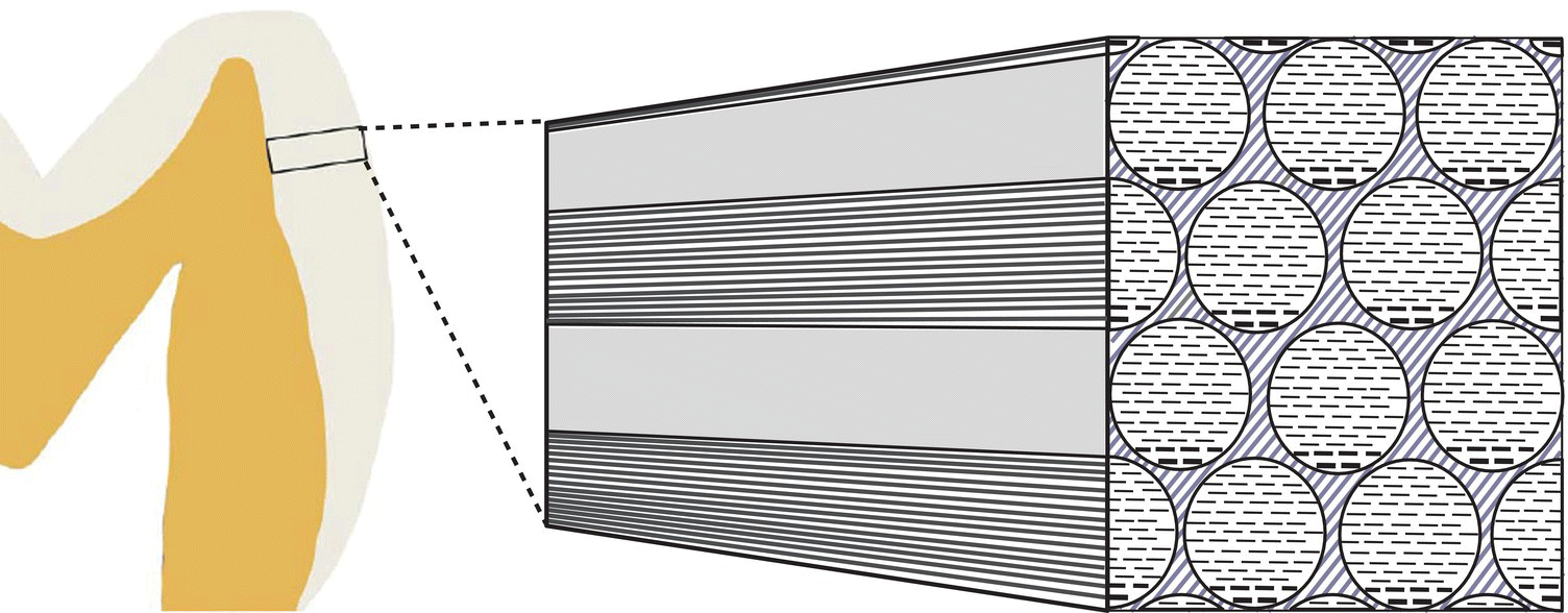
4: Enamel | Pocket Dentistry

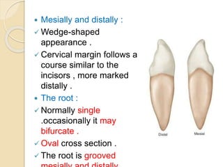
Dental anatomy
Dental anatomy

Anatomical shaping with XP 3-D Shaper and Finisher - Endodontic Practice US  - Dental Journal and Online Dental CE
Anatomical shaping with XP 3-D Shaper and Finisher - Endodontic Practice US - Dental Journal and Online Dental CE

Root Canal Anatomy | Pocket Dentistry

Longitudinal and cross sections images of the same (a) nonstained and... |  Download Scientific Diagram
Longitudinal and cross sections images of the same (a) nonstained and... | Download Scientific Diagram

Pulp Chambers and Canals (Dental Anatomy, Physiology and Occlusion) Part 3

Changed cross-sectional root canal shape for maxillary first molar from...  | Download Scientific Diagram
Changed cross-sectional root canal shape for maxillary first molar from... | Download Scientific Diagram
Dentistry and Medicine: Equipments, Instruments and Materials used in Oral  Surgery (Armamentarium)
Dentistry and Medicine: Equipments, Instruments and Materials used in Oral Surgery (Armamentarium)

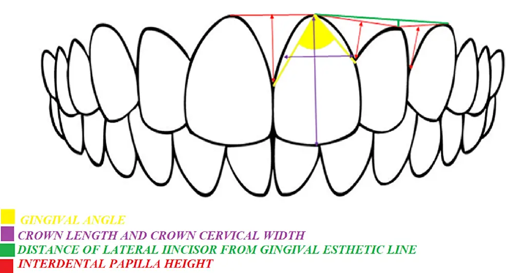
Cureus | Go Micro Aesthetics: A Cross-Sectional Study to Assess Anterior  Hard and Soft Tissue Parameters in Young Adults of Bhopal City | Article
Cureus | Go Micro Aesthetics: A Cross-Sectional Study to Assess Anterior Hard and Soft Tissue Parameters in Young Adults of Bhopal City | Article

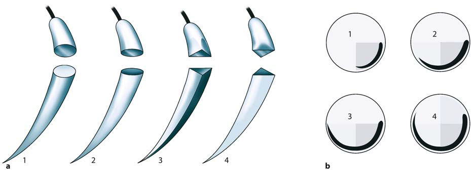
Cross-sectional root canal shape (a) and images (b) of maxillary first... |  Download Scientific Diagram
Cross-sectional root canal shape (a) and images (b) of maxillary first... | Download Scientific Diagram

The Dental cosmos. 3) Individuals, such as the Swiss type {ain Fig. 14) may  be said to lack nasaland maxillary development in compari- Fig. 26.  (Coeberg.) As of lower animals, so
The Dental cosmos. 3) Individuals, such as the Swiss type {ain Fig. 14) may be said to lack nasaland maxillary development in compari- Fig. 26. (Coeberg.) As of lower animals, so

Three-dimensional instrumentation — reaching the next level in endodontics  - Endodontic Practice US - Dental Journal and Online Dental CE
Three-dimensional instrumentation — reaching the next level in endodontics - Endodontic Practice US - Dental Journal and Online Dental CE

Crown types and cross-section outlines of the crown base at the cervix... |  Download Scientific Diagram
Crown types and cross-section outlines of the crown base at the cervix... | Download Scientific Diagram

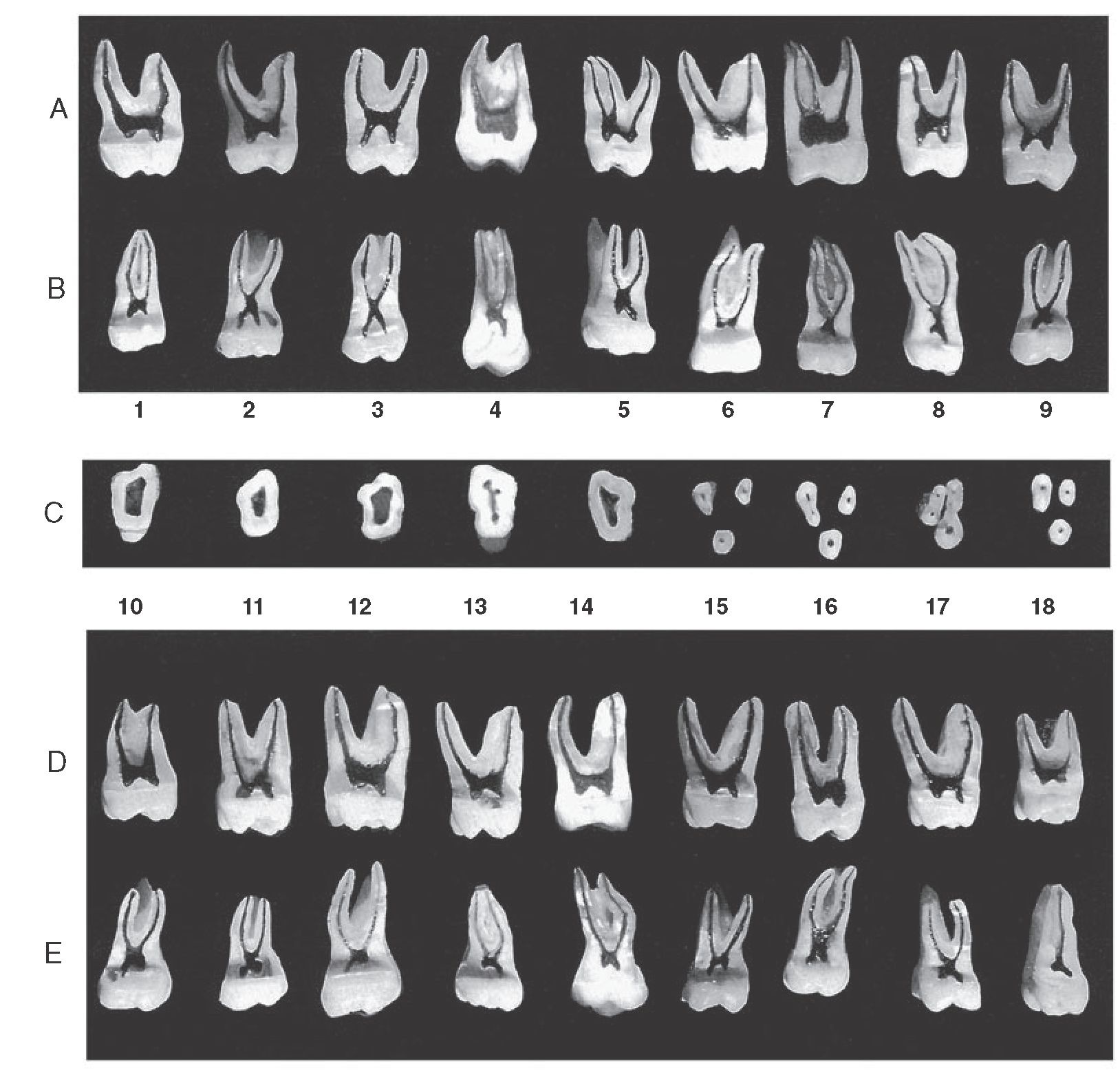
Pulp Chambers and Canals (Dental Anatomy, Physiology and Occlusion) Part 3

Crown types and cross-section outlines of the crown base at the cervix... |  Download Scientific Diagram
Crown types and cross-section outlines of the crown base at the cervix... | Download Scientific Diagram

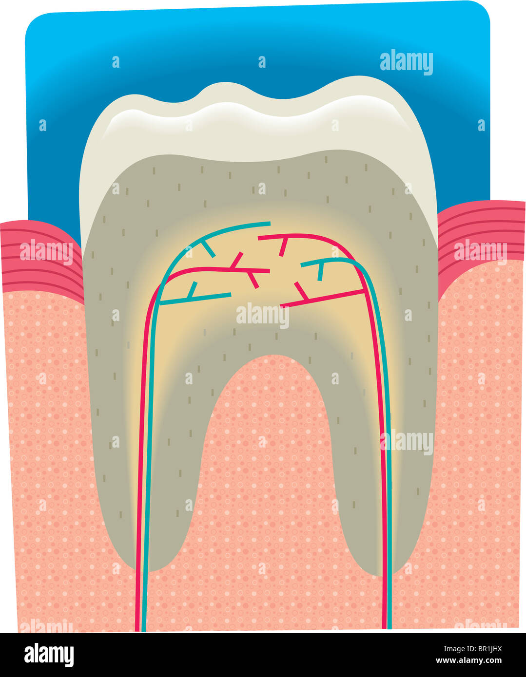
Section of a tooth hi-res stock photography and images - Alamy